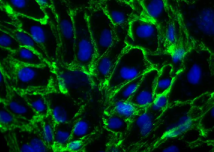
The Blood-Brain Barrier (BBB) affects the development of drugs for all pathologies (brain exposure to drugs, side effects…). In the early phases of drug development, there is a strong need for stable and fully characterized human brain endothelial cellular models that might mimic the molecular and cellular interactions between the blood and the brain in vitro.
Let’s take a look at a brain endothelial cell line with proven applications in drug discovery: the hCMEC/D3 Human Cerebral Microvascular Endothelial Cell Line.

The Human Cerebral Microvascular Endothelial Cell Line (hCMEC/D3) represents a stable, easily grown BBB cellular model in Drug Discovery and AMDE-Tox programs for:
- Drug uptake analysis
- Active transport and drug permeability
- Screening of CNS drug candidate
- Brain endothelium biology study during inflammatory stimuli or infectious diseases…
The hCMEC/D3 BBB cell line was prepared from Cerebral microvessel Endothelial Cells (CECs) by transduction with lentiviral vectors carrying the SV40 T antigen and human Telomerase Reverse Transcriptase (hTERT).
hCMEC/D3 cell line morphology and characterization
This cell line shows a spindle-shaped, elongated morphology similar to primary cultures of brain endothelial cells. It also exhibits contact inhibition at confluence when cultured on collagen type I or IV expressing brain endothelial/epithelial biomarkers together with Adherence and Tight Junction proteins and functional ABC transporters.
Interestingly, and under optimized cell culture conditions, these cells are able to maintain these unique BBB properties until the 35th subculture. When purchased via tebu-bio (cat. nr 007CLU512-A or 007CLU512-P, for academic and for-profit organisations respectively), the cell line is expected to be between the 25th-26th passage.
hCMEC/D3 cell line: a proven BBB in vitro cellular model with hundreds of international publications
If you are interested in developing cell-based assays mimicking in vitro the human blood-brain barrier, your bibliography will undoubtly bring you to the hCMEC/D3 cell line. The hCMEC/D3 cell line has been used by numerous academic and industrial organizations and is cited in 50+ international publications. A few examples:
- Maggioli E. et al. “Estrogen protects the blood–brain barrier from inflammation-induced disruption and increased lymphocyte trafficking” (2016) Brain, Behavior, and Immunity Volume 51, January 2016, Pages 212–222 – doi:10.1016/j.bbi.2015.08.020
- Mancini S. et al. “The hunt for brain Aß oligomers by peripherally circulating multi-functional nanoparticles: Potential therapeutic approach for Alzheimer’s disease” (2016) Nanomedicine: Nanotechnology, Biology and Medicine Volume 12, Issue 1, January 2016, Pages 43–52 – doi:10.1016/j.nano.2015.09.003
- Llombarta V. et al. “Characterization of secretomes from a human blood brain barrier endothelial cells in-vitro model after ischemia by stable isotope labeling with aminoacids in cell culture (SILAC)” (2016) Journal of Proteomics Volume 133, 5 February 2016, Pages 100–112 – doi:10.1016/j.jprot.2015.12.011
- Nusshold C. et al. “Assessment of electrophile damage in a human brain endothelial cell line utilizing a clickable alkyne analog of 2-chlorohexadecanal” (2016) Free Radical Biology and Medicine Volume 90, January 2016, Pages 59–74 – doi:10.1016/j.freeradbiomed.2015.11.010
- Walter F.R. et al. “A versatile lab-on-a-chip tool for modeling biological barriers” Sensors and Actuators B: Chemical (2016) Volume 222, January 2016, Pages 1209–1219 – doi:10.1016/j.snb.2015.07.110
- Weksler B. et al. “The hCMEC/D3 cell line as a model of the human blood brain barrier” (2013) Fluids and Barriers of the CNS 2013, 10:16 doi:10.1186/2045-8118-10-16
Want to know more about the human hCMEC/D3 blood-brain barrier?
You might like to get…
- An extensive list of publications describing the use of the hCMEC/D3 by clicking here.
- All morphological and phenotype characterizations of the hCMEC/D3 cell line in Dr Weksler’s initial paper that can be downloaded here.
- The experimental protocol of the hCMEC/D3 cell line here.
To further discuss your needs in the design and development of robust in vitro cellular models for studying physiopathology or drug candidates, leave a message below or subscribe to tebu-bio’s thematic eNewsletters!


2 responses
Hi Dr. Jean,
My name is Minh
Currently, I am working with hCMEC/d3. However, I have trouble with ZO-1 expression. I tried to limit the concentration of growth factors to get a high signal of ZO-1 but ZO-1 expression is still not clear.
I fixed the cell with 4% paraformaldehyde then start.
Could you please share your experience with ZO-1?
Thank you very much
Dear Tran,
Thank you for your message on our blog.
You may wish to contact the manufacturer of your antibody to gain some insight on how to optimize your protocol.
One thing to consider: is your antibody validated for PFA fixation? This may have an impact on the epitopes targeted by the antibody.
I hope this helps.
Best of luck with your project.
Kind regards,
Isabelle