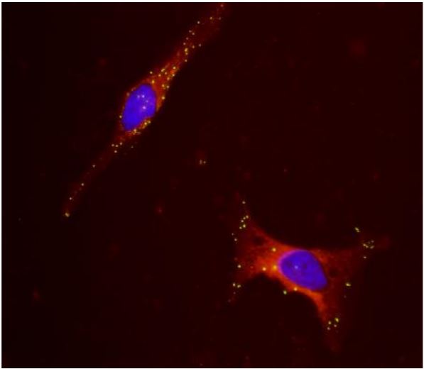
If you’re studying human pulmonary function and pathophysiology, you need access to validated, highly characterised, human airway cellular models. A large collection of these cells is available for applications in asthma, inhalation toxicology and pulmonary inflammatory response. Let’s take a look at a selection of different pulmonary cell types I would advise, to boost your in vitro research in this area.
Epithelial Cells – Bronchial & Epithelial

HBEpC & HTEpC provide an excellent model system to study all aspects of epithelial function and disease, particularly those related to airway viral infections, as well as tissue repair mechanisms, signaling changes and potential treatments relevant to lung injuries, mechanical and oxidative stress, inflammation, pulmonary diseases and smoking. HBEpC & HTEpC are obtained from healthy donors, as well as from asthma patients.
Microvascular Endothelial Cells
 Human Lung Microvascular Endothelial Cells (HLMVEC) provide a useful tool for studying various aspects of pathology and biology of the pulmonary microvasculature in vitro. HLMVEC (Cell Applications, Inc.) have been used to elucidate the therapeutic effects of Angiotensin I-converting enzyme (ACE) inhibitors, and the results revealed that they provide an additional benefit to patients by activating bradykinin B1 receptor leading to prolonged nitric oxide (NO) production in endothelial cells and inhibition of PKCϵ (Ignjatovic, 2004; Stanisavljevic, 2006).
Picture: HLMVEC in culture (A). HLMVEC immunolabeled for CD31/PECAM (green); nuclei are visualized with PI (red) (B). HLMVEC transfected with GFP plasmid DNA using the Cytofect™ Endothelial Cell Transfection Kit (C,D). Courtesy of Cell Applications, Inc.
Pulmonary Artery Smooth Muscle Cells
 HPASMC (Cell Applications, Inc). have been used to show that IL-22 promotes the growth of pulmonary vascular SMCs via a signaling mechanism that involves NADPH oxidase-dependent oxidation (Bansal, 2013). Inducers of pulmonary hypertension, characterized by thickened pulmonary arterial walls, activate expression of anti-apoptotic Bcl-xL gene via binding of GATA-4 to its promoter; the activation can be suppressed by targeting gata4 gene transcription (Suzuki, 2007). Serotonin induces growth of PHPASMC and is able to transactivate the BMP receptor in pulmonary artery SMCs, leading to activation of Smads 1/5/8 via Rho and Rho kinase pathway (Liu, 2009). HPASMC growth can be suppressed by trans-retinoic acid by inducing expression of GADD45A, a known cell growth suppressor, downstream of the retinoid acid receptors RARalpha, RARbeta, RARgamma, RXRalpha, and RXRbeta, indicating possible involvement of retinoic acid in pulmonary vascular remodeling (Preston, 2005). Treatment of HPASMC with plasma containing reduced levels of all-trans RA and 13-cis RA promoted cell growth (Day, 2009). Retinoic acid was also shown to inhibit migration of HPASMC by inhibiting PI3K/Akt-dependent reorganization of actin cytoskeleton (Day, 2006). HPASMC were also used to study effects of iron chelation on vascular remodeling and its implications for development of pulmonary hypertension as a result of ROS activity (Wong, 2012). Finally, HPASMC were used to show that antitumor drugs can selectively target remodeled pulmonary vessels, but not normal vessels (Ibragim, 2014).


