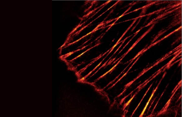
Tubulin represents one of the major cytoskeleton structures which plays an important role in cell structure, intracellular transport, and mitosis. When tubulin polymerizes to form microtubules (MTs) it initially forms proto-filaments, MTs consist of 13 protofilaments and are 25 nm in diameter, each um of MT length is composed of 1650 heterodimers. Microtubules are highly ordered fibers that have an intrinsic polarity, shown schematically in Figure 1. Tubulin can polymerize from both ends in vitro, however, the rate of polymerization is not equal. It has therefore become the convention to call the rapidly polymerizing end the plus-end of a microtubule and the slowly polymerizing end the minus-end. In vivo, the plus end of a microtubule is distal to the microtubule organizing center.


The method described here is based on the most reproducible and accurate method of determining the amount of microtubule content versus free-tubulin content in a cell population, which is to use western blot quantitation of microtubule and free-tubulin cellular fractions. The general approach is to homogenize cells in microtubule stabilization buffer, followed by centrifugation to separate the microtubules from free-tubulin pool. Then the fractions are separated by PAGE and tubulin is quantitated by western blot. The final result gives the most accurate method of determining the ratio of tubulin incorporated into the cytoskeleton versus the free-tubulin found in the cytosol. Cytoskeleton Inc. offers a ready-to-use kit which provides all components to run such assays. Exemplary results obtained with the Microtubules / Tubulin In Vivo Assay Kit are show in Fig 2. HeLa CCL-2 cells were grown to 70% confluency in DMEM/10% FBS at 37°C/5% CO2. Cells were untreated (lanes S1, P1, S2, P2) or treated with 3.3 mM of the tubulin polymerizing drug paclitaxel (i.e., taxol) for 60 min at 37°C/5% CO2 (lanes S3, P3, S4, P4). Cells were lysed and separated into supernatant (S) and pellet (P) fractions and analyzed by western blot quantitation of tubulin protein according to the Microtubules/Tubulin In Vivo Assay Kit instructions. Both gels contain the same supernatant and pellet fractions. In untreated cells, 80% of the tubulin is soluble heterodimers in the supernatant fraction (1S and 2S) while 20% is insoluble microtubules in the pellet fraction (1P and 2P). In cells treated with taxol, only 30% of the tubulin remains in the soluble heterodimer supernatant fraction (3S and 4S) while 70% is in the insoluble microtubule pellet fraction (3P and 4P). Gel 1, lane 2, and Gel 2, lane 1, contain 80 ng of purified porcine brain tubulin as a standard. Molecular weights are shown to the left of Gel 1.
Which kind of experiments can you run with the Microtubules / Tubulin In Vivo Assay Kit?
- Study the effects of pharmaceutical compounds on the ratio of tubulin to microtubules
- Study the effects of mutated cell lines versus their parent cell line for the change in ratio of tubulin to microtubules.
- Study the effects of physical alterations of environment on the ratio of tubulin to microtubules
Interested in using this method in your laboratory? Get in touch with me through the form below.
Thanks to our friends at Cytoskeleton Inc., who provided material used for this post.


