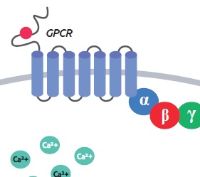
Cell-based calcium assays have been a mainstay of GPCR (G protein coupled receptor) drug discovery and signal transduction research since the mid 1990s. However, a calcium readout alone might be ambiguous, because many cellular processes produce calcium. Furthermore, it does not differentiate between signalling pathways routed through the different types of heterotrimeric G proteins. While effects on GPCRs which exhibit effects on Gs proteins lead to an elevated level of the second messaeger cAMP by activating the activity of adenylate cyclase, signalling through Gi proteins inhibits adenylate cyclase and thus leads to a decrease of cAMP. Finally signalling through Gq proteins leads to phospolipase C activation which cleaves Phosphatidylinositol 4,5-bisphosphate (PIP2) to Diacetlyglycerol (DAG) and Inositol trisphosphate (IP3) which leads to an increase in Ca2+ level (for an overview see reference 1).
Dye based methods to measure Ca2+ are very well established, whereas there are no dyes available for most of the other second messengers in these signalling pathways.
Genetically encoded fluorescent biosensors for live cell discovery
That’s why Montana Molecular developed genetically encoded fluorescent biosensors for second messengers such as cAMP, DAG, PIP2, and Ca2+, references 2, 3).
The principle of the assays is essentially based on single Red and Green Fluorescent Protein sensors, meaning the genetic information for either GFP or RFP linked to an analyte specific sensor protein domain is introduced into the cells to be investigated. Upon analyte binding to the sensor the fluorescent intensity of GFP or RFP changes which can be easily detected.
The genetic transfer is done with BacMam Vectors based on a modified baculovirus system. In mammalian cells, the baculovirus genome is silent, and it cannot replicate to produce new virus in mammalian cells. For the workflow of the assay see Figure 1.


Multiplexing in a single live-cell assay shows when an activated receptor signals through two different pathways
The availability of most of the sensors in 2 colors (GFP and RFP) allows for parallel mesurements of different second messangers. As an example Figure 2 shows how combining red cAMP sensor with green DAG sensor, and co-expressed with the Calcitonin receptor in HEK293 cells, can give you a more detailed insight into GPCR mediated pathways. While signaling through G protein Gs results in the increase of cAMP (red), Gq mediated signalling results in DAG increase (green) – meaning that both Gq and Gs pathways have been activated in this experiment.
Interested in further applications of Montana’s biosensors, especially with the goal of having a closer look at the different GPCR related signalling pathways?
 Download the poster “Development and Delivery of Robust Biosensors for Measuring Signal Transduction in Living Cells” recently presented at “Experimental Biology” (San Diego, April 2016).
Which analytes can be measured with the Montana Molecular fluorescent biosensors?
Kits are available for live cell measurement of various cellular parameters:
- cAMP,
- DAG,
- PIP2,
- Ca2+and voltage changes.
The sensors are partly available with either GFP or RFP and with different promotors optimized for different target cells (ArcLight Voltage Sensor either with CMV or synapsin promoters).
Interested? Leave your comments or requests below, I’ll be pleased to respond!
References:
(1) Stefanie L. Ritter & Randy A. Hall: Fine-tuning of GPCR activity by receptor-interacting proteins. Nature Reviews Molecular Cell Biology 10, 819-830 (2009)
(2) Paul H. Tewson et al.; A multiplexed fluorescent assay for independent second-messenger systems: Decoding GPCR activation in living cells. Journal of Biomolecular Screening 18 (7), 797-806 (2013)
(3) Paul H. Tewson et al.; New DAG and cAMP sensors optimized for live-cell assays in automated laboratories. Journal of Biomolecular Screening 21 (3), 298-305 (2016)


2 responses
It would be great if you could develop a similar readout for the G12/13 coupled Rho activation. There are no commercial assays for RhoA activation.
Dear Doug,,
thank you very much for your comment. I will inform the Montana lab about your lead.
For the time being we can indeed only offer classical RhoA activation assays such as the pulldown format (cat no.BK036) or the G-LISA format (cat. no. BK124 and BK121).
Best regards
Ali