Following the start of our recent collaboration with Phenocell, we’re pleased to be able to provide high quality Sebocytes developed from Human induced pluripotent stem cells (iPSC). Thanks to a perfectly standardized reprogramming protocol, they display lower batch to batch variability, allowing better reproducibility and accuracy of your experimental results.
Sebocytes have demonstrated their large potential to be unique tools for many life science research fields such as:
- sebocyte cell research,
- hyper- and hypo-seborrhoea-linked disorder research,
- pharmacology,
- toxicology and drug discovery, including 3D skin reconstruction.
These cryopreserved reprogrammed pluripotent stem cells are available at low passage (P2), 2.106 cells/vial format and in 3 different phototypes (Caucasian, Asian and African). Developed from highly qualified Human iPS cells, each lot is validated, with a specific certificate of analysis, for all the following Sebocyte markers and specific functions.
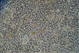
iPS-derived human Sebocyte: morphology
Phenocell’s iPS-derived human Sebocytes display the typical epithelial morphology of primary sebocytes with heterogeneity in cell size due to lipid accumulation.

Specific marker expression
Immunohistochemistry

RT-qPCR
Functional markers are strongly expressed after 5 days:
- Lipid activated transcription factor PPARγ, FADS2 (enzyme of the fatty acid biosynthesis pathway)
- Androgen receptors (AR)
- Melanocortin 5 receptor
- K7 and MUC1

Purity – Flow Cytometry analysis
KRT7 expression shows Sebocyte purity above 90%.

Function
Response to Linoleic acid

Response to testosterone

iPS-derived human Sebocyte: viability and other parameters
- Sebocytes differenciated from Human induced pluripotent stem cells: viability > 60% after thawing (Post-thaw regrowth test performed on each batch)
- Normal Karyotype
- All batches are tested negatively to mycoplasma before freezing
- Additional information regarding these Human iPSC-derived Sebocytes on tebu-bio’s dedicated web page.
Interested in these Sebocytes developed from Human induced pluripotent stem cells ?
Get in touch by leaving a comment below, I’ll be pleased to get back to you to discuss your projects and needs.


