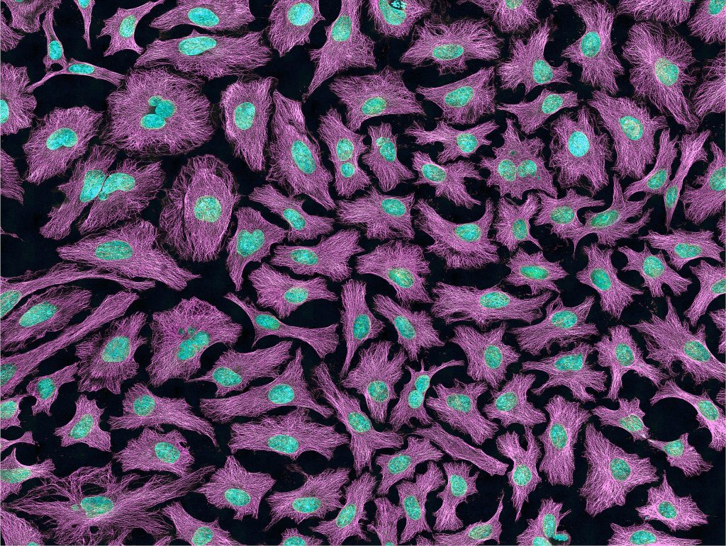
If you work in Biology, you’ve most certainly heard of HeLa cells, as they have been around for over 60 years and are some of the most extensively used cell lines in Biomedical research. But where did these cell lines come from?

In 1951, Henrietta Lacks came into John Hopkins Hospital in Baltimore, worried about a lump in her abdomen, where she was diagnosed and treated for cervical cancer (adenocarcinoma of the cervix, a particularly aggressive type of cancer). She eventually died of her cancer later that year, without knowing what her cells would help accomplish.
The surgeon treating Henrietta’s adenocarcinoma had been collecting cancerous tissue samples from patients for research lead by Dr. George Gey, Director of the Tissue Culture Laboratory at John Hopkins. His objective was to cure cancer by creating an immortalized cell line for research, in order to develop therapies and medicines.
For years, Dr. Gey and his wife Margaret (a trained surgical nurse) had been trying to cultivate human cells in vitro. All their previous attempts at growing human cells in a laboratory lead to the death of the cell cultures within a few generations. That is, until Henrietta’s tumour sample: HeLa, named after the two first letters of Henrietta and Lacks.
What’s different about HeLa cells?
There are 3 major differences between normal cells and HeLa cells:
1- HeLa cells are cancerous. The difference between normal cells and HeLa cells is most visible when you look at the chromosomes (karyotype). HeLa cells, like many tumours, have error-filled genomes, with one or more copies of many chromosomes: a normal cell contains 46 chromosomes whereas HeLa cells contain 76 to 80 (ref) total chromosomes, some of which are heavily mutated (22-25), per cell. This is due to the Human Papillomavirus (HPV), the cause of nearly all cervical cancers. HPV inserts its own DNA into host cells and the additional DNA results in the production of a p53-binding protein which inhibits it and prevents native p53 from repairing mutations and suppressing tumours, causing errors in the genome to accumulate as unchecked cellular divisions occur.
2- HeLa cells grow unusually fast, even considering their cancerous state. Indeed, HeLa cells grow easily and rapidly, doubling cellular count in only 24 hours, making them ideal for large scale testing. They grow so fast that they can contaminate and overtake other cell cultures. This is related to the fact that Henrietta Lacks had syphilis which results in an aggressive growth of cancer due to a weakened immune system. And in 2013, it was shown that the scrambled HPV genome inserted itself near the c-myc proto-oncogene in Henrietta’s genome (ref), causing its constitutive expression and the rapid replication of HeLa cells in her body.
3- HeLa cells are immortal, meaning they will divide again and again and again… This performance can be explained by the expression of an overactive telomerase that rebuilds telomeres after each division, preventing cellular aging and cellular senescence, and allowing perpetual divisions of the cells.

The HeLa cell line was the first successful attempt at immortalizing human-derived cells in vitro. In the past, researchers spent more time trying to keep cells alive than performing actual experiments. Soon after his discovery, Dr. Gey was sharing this cell line with co-workers active in cancer research and other fields, all around the world. The HeLa cell line gave them the time and the possibility to conduct repeatable experiments on human cells, without testing directly on humans. And to this day, HeLa cells have saved countless lives, and many scientific landmarks (such as cloning, gene mapping, in vitro fertilization, the polio vaccine…) have used HeLa cells and owe everything to the life and death of Henrietta Lacks.
You can find more information about Henrietta Lacks, her story, her legacy, and bioethical standards here.
HeLa cells are still widely used in research:
HeLa cell lines – robust cellular models for in vitro testing?
With all these characteristics, HeLa cells became progressively popular cellular models for life scientists willing to study mechanism of action of diseases or therapeutically active drug molecules. They have also been used to decipher cell signalling events such as DNA damage repair (ref).
Recently, HeLa cells have been used to develop cellular models in which a specific gene of interest is silenced by genome editing. Several methods are available for gene editing (ex. CRISPR-Cas 9, TALEN, stable RNAi delivery).
SilenciX® HeLa cells are tebu-bio’s knockdown (KD) cell lines based on a unique siRNA Delivery system that enables the generation of syngenic, ready-to-use and stable cellular in vitro models stably silenced for a gene of interet. This technology has already been validated in numerous scientific articles covering various biological domains and applications such as DNA Repair, Epigenetics, Ubiquitination and the cell cycle, Drug discovery (ex. synthetic lethality, personalised medicine, drug selectivity, combinatorial therapy), cell signalling and mechanism of action studies (ex. loss-of-function, Human disease model mimicking).
To know more about these SilenciX HeLa cells stably silenced please browe the technical info page, HeLa cells stably silenced: SilenciX KD cell lines, that describes the principle of the KD, the list of catalog SilenciX cell lines together with numerous scientific publications using this SilenciX technology.


3 responses
Hello,
Do you have the references still for this article? Great read by the way.
Regards,
Joshua Beeton
Dear Joshua,
Thank you for your nice comment. It was actually written by an ex colleague of mine.
The links towards the references are included in the article.
I hope this helps.
Best regards,
Isabelle
I can understand why this was diagnosed initially as a epithelioid cancer. The cytoplasm of the cells loos like squamous cell carcinoma. I understand the diagnosis was changed to adenocarcinoma at a later date. Most adenocarcinomas don’t have this type of cytoplasm. Because of that, l wondered if adenosquamous carcinoma may have been the correct diagnosis.