In a previous post I discussed 3 new unique research tools for analyzing Amyloid peptide in Alzheimer’s Disease (AD). Today, I’d like to come back to the unique properties of the Amylo-Glo® fluorescent substrate which makes Amyloid plaque staining “better, easier and faster”.
Amylo-Glo® properties
Amylo-Glo® is a newly released “Ready-to-Dilute” staining kit which contains a unique fluorescent substrate for the detection and localization of Amyloid plaques in brain tissue.
Amylo-Glo® brings superior visualization of amyloid plaques compared to other commonly used stains like Congo Red and Thioflavin S. Compatible with fresh, frozen and formalin-fixed immunohistochemistry, Amylo-Glo® is ideal for confocal microscopy including multiple labeling analysis. It’s a brighter, longer lasting, higher definition Amyloid plaque stain with:
- High fluorescent intensity
- Strong resistance to photo-bleaching
- UV channel fluorescence
Seeing is believing… the pictures speak for themselves!
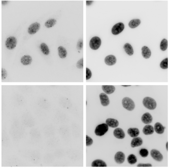
Picture 1: High magnification view of Amylo-Glo® labeled amyloid plaque from the cortex of an 11 month old AD/Tg mouse with UV illumination only. Note the high clarity, ultra low background and very fine detail available only with Biosensis Amylo-Glo® staining reagent.
Picture 2: Congo Red stain of amyloid plaque from the cortex of an 11 month old AD/Tg mouse with green light excitation. Note that while the central region of the plaque appears uniformly bright as well as some of the large fibrils, the fine details remain obscure. Also the intensity of the fluorescent emission is the weakest of all the tracers leading to early photo bleaching and making quantification and counting very difficult.
Picture 3: Thioflavin S staining of amyloid plaque from the cortex of an 11 month old AD/Tg mouse with blue light excitation. Note that while the central region of the plaque is uniformly stained, the fibrils are not; they are diffuse and cloudy. Also the background staining is relatively high, even showing some relatively faint non-specific cellular labeling as well, leading to a much more difficult plaque quantification and clarity determination.
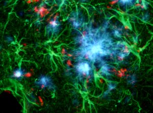
Picture 4: Simultaneous localization of Amylo-Glo® positive Amyloid plaque (blue), GFAP positive hypertrophied astrocyte (green) and activated microglia (red) in the hippocampus of the AD/Tg mouse. Combined UV, blue and green light illumination.
Want to know more?
Find out more about the Amylo-Glo® ready-to-dilute RTD™ staining reagent or contact me for further information.
[contact-form to=’philippe.fixe@tebu-bio.com’ subject=’Amylo-Glo® RTD™ staining reagent’][contact-field label=’Name’ type=’name’ required=’1’/][contact-field label=’Email’ type=’email’ required=’1’/][contact-field label=’Comment’ type=’textarea’ required=’1’/][/contact-form]


