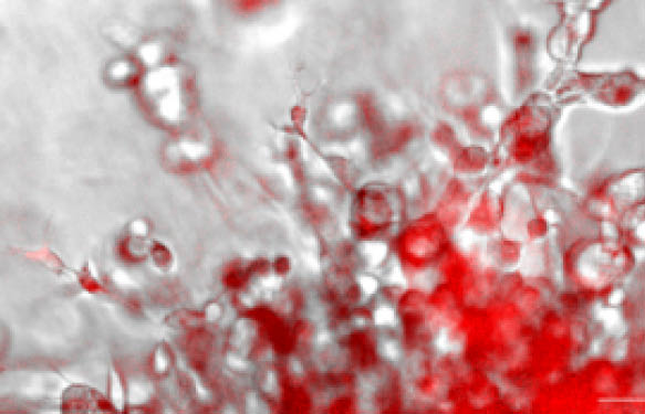
Singlet Oxygen (1O2) fluorescent detection in living cells was limited by the lack of availability of cell membrane permeable dyes. The silicon-based far-red fluorescent probe (Si-DMA) has been especially designed to monitor singlet oxygen in real-time and in living cells. Si-DMA is now commercially available for ROS studies by live-cell imaging.
Singlet oxygen (1O2) is an excited state of the dioxygen molecule. This oxidizing molecule belongs to the Reactive Oxygen Species (ROS) and is biologically involved in photodegradation and related biological events (ex. skin wrinkle with age). 1O2 high-oxidizing properties are also used as a promising anticancer treatment using photoirradiation and photosensitization in PhotoDynamic Therapies (1, 2), emphasizing the importance of precisely monitoring this molecule in cells.
The unique cell-permeable Si-DMA far-red fluorescence probe is composed of silicon-conjugated rhodamine and anthracene moieties to serve respectively as a chromophore and a 1O2 reactive site. Once in the living cells and in the presence of 1O2, the fluorescent levels of Si-DMA increases selectively due to endoperoxide formation at the anthracene moiety to selectively detect the 1O2 (see Fig.1 to 3).
Si-DMA has enabled real-time monitoring of 1O2 generation from protoporphyrin IX in mitochondria with 5-aminolevulinic acid (5-ALA), a precursor of heme (see Fig. 4 in HeLa cells).
Si-DMA for Mitochondrial Singlet Oxygen Imaging (Cat. nr MT05-10) complements the series of other cell-permeable proprietary “silicon-rhodamine” fluorophore (eg. SpiroChrome) commonly used to selectively monitor in live cell imaging tubulin (SiR-tubulin), actin (SiR-Actin), and DNA (SiR-DNA).
What about you?
Working on Reactive Oxygen Species or Mitochondria and live-cell imaging? Which fluorescent dyes are you using, or would you be interested in using in the future?
Share your comments, thoughts and questions below!
Sources:
1) T. Majima, S. Kim, T. Tachikawa, M. Fujitsuka, “Far-Red Fluorescence Probe for Monitoring Singlet Oxygen during Photodyanamic Therapy”, J. Am. Chem. Soc., 2014, 136 (33), 11707-11715.
2) T. Majima, S. Kim, T. Tachikawa, M. Fujitsuka, WO 2015194606, A1, (23, December, 2015).
 Want to learn more about tools like this?
Subscribe to thematic newsletters on your favourite research topics.


