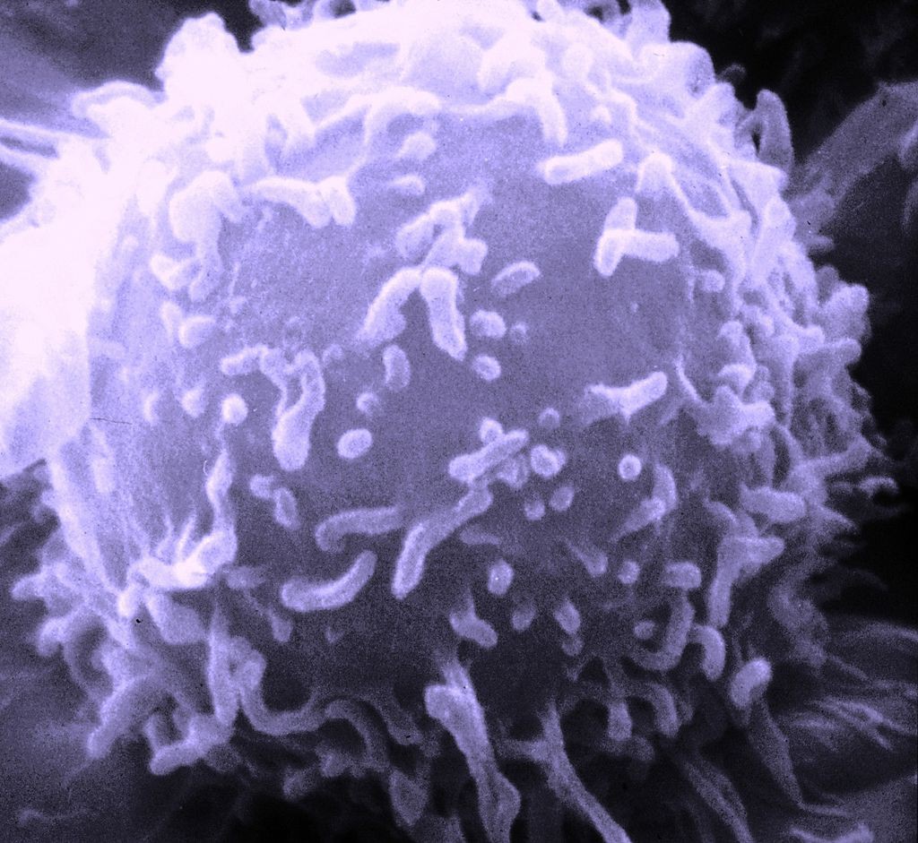
Classically, it was known that CD4+ T cells, upon activation and expansion, develop into different T helper cell subsets with different cytokine profiles and distinct effector functions to effect the immune response. Until some years ago, T cells were divided into Th1 or Th2 cells, depending on the cytokines they produce (1).
However, a third subset of IL-17-producing effector T helper cells, called Th17 cells, was discovered and characterised. Th17 cells produce several factors that can be quantified with one single multiplex immunoassay (e.g. IL-17, IL-17F, and IL-22). Th17 cells seem to induce a massive tissue reaction owing to the broad distribution of the IL-17 and IL-22 receptors.
Th17 cells also secrete IL-21 to communicate with the cells of the immune system. The differentiation factors (TGF-β plus IL-6 or IL-21), the growth and stabilization factor (IL-23), and the transcription factors (STAT3, RORγt, and RORα) involved in the development of Th17 cells have also been identified. Th17 cells thereby seem to be mainly involved in clearing pathogens during host defense reactions and in inducing tissue inflammation in autoimmune disease.
Let’s focus on the role of TGF-β. Though it has been shown to positively regulate the development of murine T helper type 17 (Th17) cells, which of the intracellular signaling pathways are involved is controversial. A recent publication using protein antibody arrays found an increase of expression and phosphorylation of the following Smad-independent signaling molecules in Th17-polarized wild-type T cells: AKT1(Tyr474), AKT2 (Ser474), ERK1-p44/42 MAPK(Tyr204), mTOR(Thr2446), p38 MAPK(Thr180), Rac1/cdc42(Ser71), SAPK/JNK(Tyr185) and SP1(Thr739) (2).
Discovery of so many signaling molecules was only possible as the antibody arrays allowed the screen dozens (or even hundreds!) of targets in a simultaneous way. Probably, if the research had involved picking different targets and doing individual WBs, there would have been a byass on what markers to study, and some of the ones mentioned above may never have been detected.
Th17 cells also have a role in tumour microenvironment (TME). It has been found that the Th17 cell survival factor, IL-23, is overexpressed in tumor tissues isolated from mice and human breast cancer patients. It has been indicated that tumor-secreted PGE2 induces IL-23 production in the TME, leading to Th17 cell expansion. This inductive effect of PGE2 is mediated through cAMP/PKA signaling transduction pathway (3).
For further updates on effector molecules in the Th17 pathway, don’t hesitate to suscribe to our blog!
References
1.- Korn, R. et al (2009). Annual Review of Immunology. Vol. 27: 485-517. DOI: 10.1146/annurev.immunol.021908.132710.
2.- Hasan, M. et al (2015). Immunol Cell Biol. 2015 Aug;93(7):662-72. doi: 10.1038/icb.2015.21.
3.- Qian, X. et al (2013). J Immunol. 2013 Jun 1;190(11):5894-902. doi: 10.4049/jimmunol.1203141.


