In this post, I’d like to take a look at the current understanding of tubulin PTMs, that include tyrosination/detyrosination, Δ2-tubulin formation, acetylation, phosphorylation, ubiquitination, glutamylation, and glycylation. This is inspired by contribution provided by Cytoskeleton Inc., who are experts in this domain.
Post-translational modifications (PTMs) are highly dynamic and often reversible processes where a protein’s functional properties are altered by addition of a chemical group or another protein to its amino acid residues. As a major cytoskeletal protein with roles in cell development, growth, motility, and intracellular trafficking, tubulin and microtubules (MTs) are a major substrate for PTMs. Tubulin PTMs usually occur post-polymerization and preferentially on the a/b tubulin heterodimers of stable (vs dynamic) MTs1-3.
(De)tyrosination and Δ2-tubulin generation
Tyrosination of brain tubulin was one of the earliest reported tubulin PTMs, first described in 19744. Tyrosination and detyrosination are reversible PTMs (see Fig. 1) occurring on most alpha-tubulin subunits, but not on beta-tubulin. MTs, but not tubulin heterodimers, undergo detyrosination where the gene-encoded C-termini amino acid tyrosine is proteolytically removed. Detyrosination is associated with MT stability as long-lived MTs in many different types of cells lack tyrosination. However, detyrosination itself does not confer stability to the MTs5.

Interestingly, exogenous stabilization of MTs with taxol increases the level of detyrosination1. Once detyrosinated, tubulin can be further converted into Δ2-tubulin by the irreversible removal of the penultimate glutamate residue on alpha-tubulin (see Fig. 1). Δ2-tubulin cannot be re-tyrosinated. Δ2-tubulin is also a characteristic of stable MTs such as those found in neurons, centrosomes, and primary cilia6. Δ2-tubulin is particularly abundant in brain (representing 35% of total tubulin), especially in differentiated neurons6. In MTs that have only been detyrosinated, the alpha-tubulin C-termini are re-tyrosinated, but only once the MTs have depolymerized into tubulin heterodimers7. MTs in neuronal axons are prime examples of a mixed population of detyrosinated and tyrosinated MTs. MTs in the outgrowing neuronal axon are composed of a labile and dynamic domain at the plus (distal) end that is enriched in tyrosinated tubulin and nearly devoid of detyrosinated tubulin. Conversely, at the proximal end of the axon, there is a stable domain enriched in detyrosinated tubulin that is nearly devoid of tyrosinated tubulin. These observations led to the hypothesis that MT dynamics differ throughout the axon and growth cone of neurons1-3,7.
The tyrosination/detyrosination PTMs are believed to be associated with at least two MT-associated processes which may be inter-related. One is stabilization of MTs and the second is mediating the interaction of MTs with microtubule-associated proteins (MAPs). In vitro studies with purified tubulin detect no effects of detyrosination on polymerization dynamics8-10.
Acetylation

The tubulin PTM of acetylation was first reported in the early 1980s. Acetylation of the epsilon amino group of lysine at position 40 (Lys40) occurs on alpha-tubulins of assembled MTs including those of cytoplasmic, spindle, centriolar, and axonemal origins (spanning algae to mammals)1-3 (see Fig. 2). The acetylation of Lys40 is only found on alpha tubulins and is considered the primary site of tubulin acetylation. Similar to detyrosination, acetylation is considered a marker of MT age (MTs that are more stable and resistant to turnover). Recently, other lysine residues in alpha-tubulin and beta-tubulin have been reported to be targets for acetylation based on proteomic analyses. Some of these residues are on the outer surface of the MT, as opposed to Lys40, which is in the MT lumen2,11. Acetylated tubulin is considered a marker of stable MTs resistant to nocodazole, although acetylation itself does not cause MT stabilization12. Under depolymerizing conditions (nocodazole or colchicine, but not cold), acetylated MTs are more stable than non-acetylated MTs. When stabilized by taxol, MTs become acetylated, suggesting MT stability occurs independently of acetylation13,14. Acetylation does not affect tubulin polymerization or depolymerization15. The dogma that acetylation is restricted to stable MTs has been revised in recent years and now it is known that tubulin acetylation is also found on dynamic MTs15. Particular cell subpopulations that do not have stable MTs can still undergo MT acetylation. In mature neurons, acetylated MTs are non-uniformly distributed with enrichment in the proximal region of axons and present at decreased levels in the cell body, dendrite, and growth cone. Conversely, in young neurons, acetylated MTs are in the proximal regions of the neurites and cell body undergoing neurite outgrowth7,15,16.
Acetylated MTs are found in many different types of cells and structures, including flagella of algae, neurons, primary cilia of mammalian cells, and spindle MTs, but the function of acetylation in all of these populations is still very unclear15. Recent studies suggest that tubulin acetylation has multiple functions in cells, including ciliogenesis in mammals as well as a role in neuronal neurite development and MAP binding. In neurons, acetylation by the -acetyltransferase ARD1-NAT1 complex was shown to be important for dendrite extension and arborization. Likewise, inhibition of the Elongator complex results in impaired neuronal migration and branching1-3,17. Creppe et al.17 showed that overexpression of a non-acetylatable mutant alpha tubulin in cortical neurons impairs migration and branching of neurite projections. Also, the velocity of BDNF-associated vesicles was reduced as they moved along MTs1. Thus, acetylation in neurons could be relevant for migration, differentiation, growth, and synaptic targeting of proteins.
Phosphorylation

Phosphorylation of tubulin was first reported in the early 1970s by multiple research groups18-20. In the past 4 decades, our understanding of the phosphorylation of tubulin, the sites affected, and the physiological relevance remains severely limited. While most tubulin PTMs act on tubulin subunits already incorporated into MTs, phosphorylation can act on both tubulin heterodimers and polymers. One of the tubulin residues targeted for phosphorylation is Serine172 (Ser172). In the case of Ser172, phosphorylation only occurs on beta-tubulin21 (see Fig. 3). Besides phosphoserine 172, other serine as well as tyrosine residues on tubulin alpha/beta heterodimers (and MTs) are presumed targets of phosphorylation22-28.
The physiological relevance of tubulin phosphorylation remains an active area of research with studies suggesting a role for phosphorylation in regulating polymerization, both positively and negatively. An early functional study found that phosphorylation of beta-tubulin is increased by serum deprivation-induced cell differentiation. The level of phosphorylation is also influenced by the state of MT polymerization as taxol stimulates phosphorylation while nocodazole decreases it29. Although this study29 did not report which residues were phosphorylated, more recent studies have focused on tyrosine and serine residues. Laurent et al.27 found that the non-receptor tyrosine kinase Fes not only phosphorylates tubulin, but also promotes tubulin polymerization. However, the exact role phosphorylation might have in the Fes-mediated enhancement of polymerization is unclear since the stoichiometric phosphorylation of tubulin was not required for increased MT assembly27.
Ubiquitination
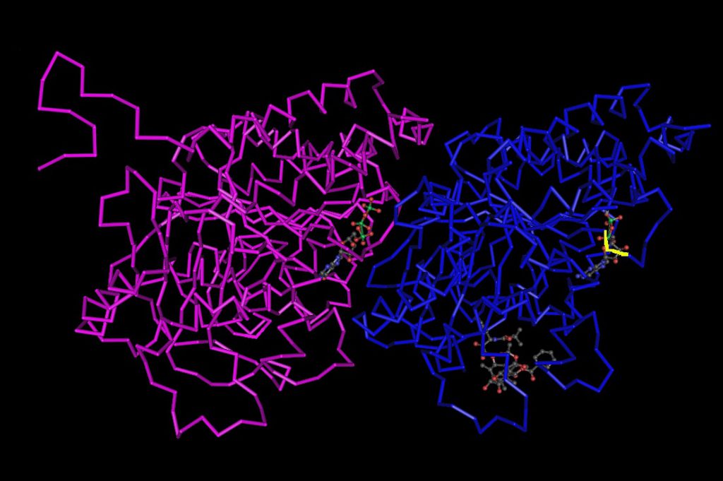
Within the last two decades, researchers have reported that another tubulin PTM is ubiquitination and recent research efforts are focused on examining how ubiquitination affects tubulin polymerization and function. Tubulin heterodimers that comprise the MTs of various subpopulations (mitotic spindle, neuronal projections, centrioles/basal bodies, and axenomes) are the target of various ubiquitin ligases (see Fig. 4).

The physiological relevance of tubulin ubiquitination remains under investigation, but it is known that gamma-tubulin is ubiquitinated by the ubiquitin ligase BRCA1, a modification required for tubulin nucleation and normal centrosome function32-34. Ubiquitination has also been implicated in α/β-tubulin turnover and ubiquitination of α/β-tubulin has been observed in various cellular processes30,35-37. UCH L1 is a multi-functional protein with ubiquitin ligase activity in vitro31. Recently, UCH L1 was identified as a tubulin-interacting protein by mass spectrometry, and a UCH L1 mutation associated with Parkinson’s disease as well as carbonyl-modified UCH L1 promotes tubulin polymerization38.
Glutamylation and Glycylation

Polyglutamylation and polyglycylation are the addition of one or more glutamate or glycine residues, respectively39,40, to a/b tubulin heterodimers. These modifications, known as polymodifications, were discovered in the early 1990s and are often discussed together as the same glutamate residues are the substrate for these PTMs (see Fig. 5). The MTs that comprise the mitotic spindle, neuronal projections, centrioles/basal bodies, and axenomes undergo polyglutamylation. Conversely, polyglycylation occurs mainly on MTs of the axoneme. The axoneme is the cytoskeletal structure that comprises flagella and cilia. Cilia and flagella are evolutionarily conserved MT-based organelles protruding from the cell surface that perform dynein-driven beating for cell locomotion or flow generation. They also play important roles as cellular antennae, allowing cells to respond to external stimuli. Flagella are also found on cells with a primary role in human reproduction41-43. Recently, abnormal formation or function of cilia has been linked to a broad range of human genetic disorders termed ciliopathies41. Given the prominent roles that tubulin glutamylation and glycylation play in cilia function (see below), these polymodifications may be involved in a myriad of human diseases.
Despite targeting the same glutamate residues, these polymodifications have unique attributes. While both tubulin subunits undergo both polymodifications, the extent of the modification is spatially regulated. For example, in superior cervical ganglion neurons, tubulin glutamylation is abundant on beta-tubulin in somatodendritic regions, while alpha-tubulin is glutamylated to a greater extent in axons44. Furthermore, glutamylation of alpha-tubulin is abundant in young neurons whereas glutamylation of beta-tubulin increases during post-natal development45. In vivo, the length of the polymodification side chains correlates with the type of MT. For example, in ciliates, the side chains are longer in axonemes compared to MTs in the cell body1,46,47. Also, in vivo competition between the polymodifications is reported, likely due to the fact that both modifications occur on the same substrate: glutamate residues in the CTT42,43. This has led researchers to speculate that the two polymodifications negatively regulate each other.
Both polymodifications occur on the CTT which is where structural and motor MAPs bind48, suggesting that these PTMs could regulate such binding to confer MT functional diversity1-3. Indeed, recent in vitro studies demonstrate that tubulin polyglutamylation can modulate the binding of structural and motor MAPs to MTs, which could serve as a means of controlling the functional specificity of MT subpopulations49,50-52.
Tools to study PTMs of tubulin
To provide researchers with the tools to study tubulin PTMs, Cytoskeleton Inc offers purified tubulin proteins, PTM antibodies, and activation assays. Cytoskeleton has purified mammalian brain tubulins as well as tubulin from HeLa and MCF-7 cancer cell lines. These purified tubulins can be incubated with modifiers such as kinases, ubiquitin ligases, tyrosine ligases, glutamylases, glycylases, and HDAC6, as well as the enzymes that reverse PTMs. To detect tubulin PTMs, you can choose from a set of PTMtrue antibodies:
- Anti-acetyl lysine antibody
- Monoclonal anti-phosphotyrosine antibody
- Anti-ubiquitin antibody
- Anti SUMO-2/3 Mouse Monoclonal Antibody
- Pan-anti-tubulin antibody that detects various isoforms of tubulin from multiple species and labels tubulin independent of PTMs
To study changes in polymerization of tubulin, you can use polymerization assay kits with different types of tubulin based on the levels of MAPs present.
Thanks to our friends from Cytoskeleton Inc, who provided the information summarized in this blog!
Interested in these tools? Ask your questions and feel free to leave your comments below!
Subscribe to thematic newsletters on your favourite research topics
References:
1. Wloga D. and Gaertig J. (2010). J. Cell Sci. 123, 3447-3455.
2. Janke C. and Kneussel M. (2010). Trends Neurosci. 33, 362-372.
3. Hammond J., Cai D., and Verhey K.J. (2008). Curr. Opin. Cell Biol. 20, 71-76.
4. Barra H.S., Arce C.A., Rodriguez J.A., and Caputto R. (1974). Biochem. Biophys. Res. Commun. 60, 1384-1390.
5. Khawaja S., Gundersen G.G., and Bulinksi J.C. (1988). J. Cell Biol. 106, 141-149.
6. Paturle- Lafanechère L., Manier M., Trigault N., Pirollet F., Mazarguil H., et al. (1994). J. Cell Sci. 107, 1529-1543.
7. Fukushima N., Furuta D., Hidaka Y., Moriyama R., and Tsujiuchi T. (2009). J. Neurochem. 109, 683-693.
8. Maruta W.T., Greer K., and Rosenbaum J.L. (1986). J. Cell Biol. 103, 571-579.
9. Paturle L., Wehland J., Margolis R.L., and Job D. (1989). Biochemistry. 28, 2698-2704.
10. Webster D.R., Wehland J., Weber K., and Borisy G.G. (1990). J. Cell Biol. 111, 113-122.
11. Choudhary C., Kumar C., Gnad F., Nielsen M.L., Rehman M., et al. (2009). Science. 325, 834-840.
12. Palazzo A., Ackerman B., and Gundersen G.G. (2003). Nature. 421, 230.
13. Piperno G., LeDizet M., and Chang X.J. (1987). J. Cell Biol. 104, 289-302.
14. Wilson P.J. and Forer A. (1997). Cell. Motil. Cytoskeleton. 37, 20-32.
15. Perdiz D., Mackeh R., Pous C., and Baillet A. (2011). Cell. Signal. 23, 763-771.
16. Witte H., Neukirchen D., and Bradke F. (2008). J. Cell Biol. 180, 619-632.
17. Creppe C., Malinouskaya L., Volvert M.L., Gillard M., Close P. et al. (2009). Cell. 136, 551-564.
18. Goodman D.P.B., Rasmussen H., DiBella F., and Guthrow C.E. Jr. (1970). Proc. Natl. Acad. Sci. U.S.A. 67, 652-659.
19. Murray A.W. and Froscio M. (1971). Biochem. Biophys. Res. Commun. 44, 1089-1095.
20. Eipper B.A. (1972). Proc. Natl. Acad. Sci. U.S.A. 69, 2283-2287.
21. Fourest-Lieuvin A., Peris L., Gache V., Garcia-Saez I., Juillan-Binard C., et al. (2006). Mol. Biol. Cell. 17, 1041-1050.
22. Wandosell F., Serrano L., and Avila J. (1987). J. Biol. Chem. 262, 8268-8273.
23. Matten W.T., Aubry M., West J., and Maness P.F. (1990). J. Cell Biol. 111, 1959-1970.
24. Faruki S., Geahlen R.L., and Asai D.J. (2000). J. Cell Sci. 113, 2557-2565.
25. Vogel J., Drapkin B., Oomen J., Beach D., Bloom K., et al. (2001). Dev. Cell. 1, 621–631.
26. Mitsopoulos C., Zihni C., Garg R., Ridley A.J., and Morris J.D.H. (2003). J. Biol. Chem. 278, 18085-18091.
27. Laurent C.E., Delfino F.J., Chen H.Y., and Smithgall T.E. (2004). Mol. Cell. Biol. 24, 9351-9358.
28. Ma X. and Sayeski P.P. (2007). Biochemistry. 46, 7153-7162.
29. Gard D.L. and Kirschner M.W. (1985). J. Cell Biol. 100, 764-774.
30. Ren Y., Zhao J., and Feng J. (2003). J. Neurosci. 23, 3316-3324.
31. Liu Y., Fallon L., Lashuel H.A., Liu Z., and Lansbury P.T. Jr. (2002). Cell. 111, 209-218.
32. Starita L.M., Machida Y., Sankaran S., Elias J.E., Griffin K. et al. (2004). Mol. Cell. Biol. 24, 8457-8466.
33. Sankaran S., Crone D.E., Palazzo R.E., and Parvin J.D. (2007). Cancer Biol. Ther. 6, 1853-1857.
34. Parvin J.D. and Sankaran S. (2006). Cell Cycle. 5, 1946-1950.
35. Huang K., Diener D.R., and Rosenbaum J.L. (2009). J. Cell Biol. 186, 601-613.
36. Thompson W.E., Ramalho-Santos J., and Sutovsky P. (2003). Biol. Reprod. 69, 254-260.
37. Iqbal K, Grundke-Iqbal I. (1991). Mol. Neurobiol. 5, 399-410.
38. Kabuta T., Setsuie R., Mitsui T., Kinugawa A., Sakurai M. et al. (2008). Hum. Mol. Genet. 17, 1482-1496.
39. Edde B., Rossier J., Le Caer J.-P., Desbruyeres E., Gros F., et al. (1990). Science. 247, 83-85.
40. Redeker V., Levilliers N., Schmitter J.-M., Le Caer J.-P., Rossier J., et al. (1994). Science. 266, 1688-1691.
41. Waters A.M. and Beales P.L. (2011). Pediatr. Nephrol. 26, 1039-1056.
42. Wloga D., Webster D., Rogowski K., Bre M.H., Levilliers N., et al. (2009). Dev. Cell. 16, 867-876.
43. Rogowski K., Juge F., van Dijk J., Wloga D., Strub J.-M., et al. (2009). Cell. 137, 1076-1087.
44. Ikegami K., Mukai M., Tsuchida J., Heier R.L., Macgregor G.R., et al. (2006). J. Biol. Chem. 281, 30707-30716.
45. Audebert S., Koulakoff A., Berwald-Netter Y., Gros F., Denoulet P., et al. (1994). J. Cell Sci. 107, 2313-2322.
46. Iftode F., Clerot J.C., Levilliers N. and Bre M.H. (2000). Biol. Cell. 92, 615-628.
47. Wloga D., Rogowski K., Sharma N., Dijk J.V. Janke C. et al. (2008). Eukaryotic Cell. 7, 1362-1372.
48. Nogales E. (2000). Annu. Rev. Biochem. 69, 277-302.
49. Boucher D., Larcher J.C., Gros F., and Denoulet P. (1994). Biochemistry. 33, 12471-12477.
50. Bonnet C., Boucher D., Lazereg S., Pedrotti B., Islam K., et al. (2001). J. Biol. Chem. 276, 12839-12848.
51. Larcher J.C., Boucher D., Lazereg S., Gros F., and Denoulet P. (1996). J. Biol. Chem. 271, 22117-22124.
52. Ikegami K., Heier R.L., Taruishi M., Takagi H., Mukai M., et al. (2007). Proc. Natl. Acad. Sci. U.S.A. 104, 3213-3218.


