 PD-1:PD:-L1 immune checkpoint pathway targeting immunotherapies have shown great potential for many cancer patients. Recently, The FDA has granted accelerated approval to the immunotherapy drug pembrolizumab (Keytruda® – humanized monoclonal IgG4 antibody against human cell surface receptor PD-1) for use in some patients with advanced gastric stomach cancer (see NIH-NCI news). However, response to these treatments is not guaranteed for each patient. With the complexity of the immune system and tumour microenvironment, providing more physiological outcomes for immunotherapies is necessary. Biochemical assays alone cannot consider the functionality of the whole cell signalling pathway. Complementing them with cell-based assays provides a comprehensive approach for identifying and developing new and improved immunotherapy treatments. Cellular line engineering has simplified and accelerated the development of such immunoassays. Targets have been chosen from both immune activators and suppressors with the aim of obtaining precise control of the immune system.
BPS Bioscience have engineered 6 cellular lines, turning them into cell-based reporter assays for Human Immune Checkpoint research. I’d like to present a summary of how each cellular line functions.
Cellular lines reporting Immune Inhibition:
PD-1:PDL-1 – Programmed Cell Death 1
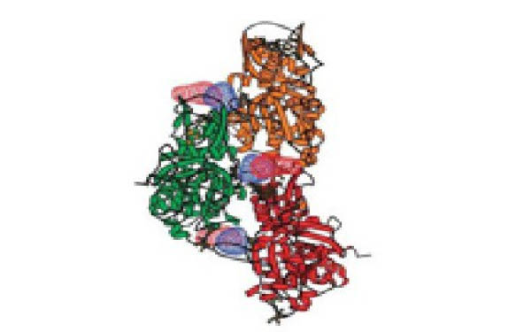
 PD-1, a receptor expressed on the surface of activated T cells, binds to its ligand PD-L1 and negatively regulates immune responses. PD-1 ligands (PD-L1) are found on most cancer cells, allowing them to dodge immune surveillance through its interaction with PD-1. Jurkat cells constitutively expressing PD-1 and the firefly luciferase gene controlled by NFAT response elements interact with cells constitutively expressing PD-L1 and an engineered TCR-activator (fig.1). The PD-1:PD-L1 interaction between these cells prevents luciferase expression. The anti-PD-1 antibody inhibits PD-1:PD-L1 interaction allowing luciferase expression and production of reporter signal (fig.2). The in vitro biochemical assay of protein-protein interaction between the extracellular domains of PD-1 and PD-L1 is monitored by TR-FRET. BMS202 inhibits the PD-1:PD-L1 interaction by stabilizing PD-L1:PD-L1 dimerization (fig.3 & fig.4).
TIGIT:CD155 – T-cell immunoreceptor with Ig and ITIM domains (TIGIT)
 TIGIT is a co-inhibitory receptor expressed in Natural Killer cells and activated regulatory T cells. Interaction with CD155 on antigen presenting cells suppresses NF-ƙB and NFAT-TCR signalling. These interactions block T cell proliferation and cytokine production. CHO-K1 cells constitutively expressing CD155 and a TCR-activator (#60548) interact with Jurkat cells that express the firefly luciferase gene controlled by NFAT response elements and constitutively express TIGIT. The TIGIT:CD155 interaction inhibits luciferase expression (#60538) (fig.5). Anti-TIGIT antibody inhibits the TIGIT:CD155 interaction (fig.6), allowing the production of the reporter signal. The Alphalisa™ proximity assay is used to monitor protein-protein interaction between the purified extracellular domains of TIGIT:CD155 (#72029). The Anti-TIGIT antibody inhibits the TIGIT:CD155 interaction (fig.7 & fig.8).
LAG3:MHCII – Lymphocyte-Activation Gene 3 (LAG3)
 LAG3 is expressed on activated T cells, natural killer cells, B cells, and APCs. Its interaction with the Major Histocompatibility Complex (MHC) class II molecules negatively regulates cellular proliferation, activation, and homeostasis of T cells, and plays a role in Treg suppressive function. Jurkat cells constitutively expressing LAG3 and expressing firefly luciferase under the control of inducible NFAT response elements are co-cultured with RAJI cells, and incubated with Superantigen (fig.21). Anti-LAG3 antibody inhibits LAG3:MHCII interaction, allowing luciferase expression and production of reporter signal (fig. 22).
Cellular lines reporting Immune Activation :
GITR:GITRL – Glucocorticoid-induced TNFR Related Protein (GITR)
 GITR is expressed in various cells including T cells, Natural Killer cells and Antigen-Presenting Cells (APCs). GITRL is expressed primarily by APCs and its interaction with GITR contributes to the activation of the immune system. Jurkat cells constitutively expressing GITR and a luciferase gene controlled by NF-ƙB response elements are treated with soluble GITRL protein (#71190). The GITR:GITRL interaction stimulates luciferase expression (fig.9). The anti-GITR antibody inhibits GITR:GITRL interaction, precluding the production of the reporter signal (fig.10). The TR-FRET proximity assay is used to monitor the interaction between the purified, extracellular domains of GITR and GITRL. The anti-GITR antibody inhibits the GITR:GITRL interaction (fig.11 & fig.12).
OX40:OX40L – OX40 (CD40, TNFRSF4, Bp50)
 Similar to the GITR assay, HEK293 cells constitutively expressing OX40 and an engineered luciferase gene controlled by NF-ƙB response elements are treated with soluble OX40L protein (#71185). The OX40:OX40L interaction stimulates luciferase expression (fig.13 & fig. 14). This chemiluminescent, direct assay is used to monitor protein-protein interaction between the purified, extracellular domains of OX40 and OX40L. Unlabelled OX40L inhibits the OX40:OX40L(Labeled) interaction (fig.15 & fig.16).
In a recent study in mice model, researchers have suggested that for treatment combining two immunotherapy drugs againt 2 immune-checkpoints (PD-1 Inhibitors & OX40 Agonists), the timing and sequence of the drugs’ administration are critical to the treatment’s efficacy and safety (see “Timing and Sequence Critical for Immunotherapy Combination” – NIH-NCI blog).
CD40:CD40L – Cluster of Differentiation 40 (CD40, TNFRSF5, Bap50)
 CD40 is found on B lymphocytes and APCs and is over-expressed in a variety of carcinoma cells. Interaction with CD40 ligand (CD40L, CD154) on CD4+ T helper lymphocytes triggers the expression of pro-inflammatory cytokines. HEK293 cells that constitutively express CD40 and an engineered luciferase gene controlled by NF-ƙB response elements are treated with soluble CD40L protein (#71191). The CD40:CD40L interaction stimulates luciferase expression (fig.17). Anti-CD40 antibody inhibits the CD40:CD40L interaction, blocking luciferase expression and the production of reporter signal (fig.18). The chemiluminescent, direct assay is used to monitor the protein-protein interaction between purified, extracellular domains of CD40 and CD40L. The anti-CD40L antibody inhibits the CD40:CD40L interaction (fig.19 & fig.20).
To perform all these cell based assays in the best conditions, I invite you to explore the many related products that are available, such as growth and thawing media, luciferase buffer, control antibodies, as well as control cellular lines.
Regarding Immunotherapy and cancer research, you might also be interested in other products such as proteins, antibodies, inhibitor/activator molecules, chemical compounds, and cellular lines easily accessible through this search engine.
These cell lines are available to test for three months at 50% of their price, after which you can decide if you wish to fully purchase or not. Contact your local tebu-bio office to learn more.
Finally, if you’re considering outsourcing cellular line engineering services, recombinant protein production or any other (part) of your projects, you can always call upon a broad range of Custom services, performed in European based laboratories.


