
G Protein Coupled Receptors (GPCR) and ion channels are important targets in drug development. Both target classes represent membrane proteins which require convenient and robust cell-based assays for screening campaigns. Usually used as readout of such screenings, intracellular Calcium concentration implies the use of Ca2+ indicators able to meet demanding expectations of researchers engaged in assay development and screenings.
Measurement of intracellular Calcium based on fluorescent dyes
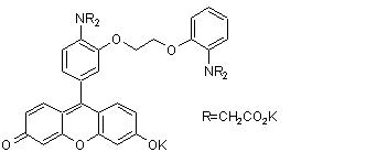 Chemical structure of Fluo-8
Commonly used tools to directly measure Calcium are Ca2+ chelators – in this case fluorescent dyes which directly bind Ca2+. Upon binding, the fluoresence intensity is significantly increased and can be easily measured by fluorimetric instruments. Classical dyes are Fura-2, Indo-1, Fluo-3, Fluo-4.
Fluo-8 (chemical structure, see Fig 1) can be considered as the state-of the-art indicator for measuring intracellular Ca2+ concentration, as it has 3 main benefits over classical fluorescent dyes:
- Twice as bright as Fluo-4 and four time brighter than Fluo-3
- Loading of the cell can be conducted at room temperature, meaning under less harsh conditions than with Fluo-3 and Fluo-4 which require 37°C for optimal loading.
- As especially HTS screening requires homogeneous assays (assays not requiring any wash steps to be HTS compatible), a Fluo-8 no wash kit is available as well.
 Fluo-8 dye is twice brigther than Fluo-4.
Interested in testing our Fluo-8?
Just contact me with the form below.
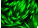
 Rat embryo fibroblasts before and after stimulation with 2 µM Ionomycin and loaded with Fluo-3, Fluo-4, or Fluo-8.