

An Insight from the Tebubio Team
Discover how small-molecule probes revolutionise cell biology by specifically labelling peroxisomes for advanced imaging and research.
Get the opportunity to access cutting-edge tools to enhance your peroxisome studies.
Streamlining Peroxisome Imaging: A New Era with Peroxi_SPY Probes
In the world of scientific research, the need for efficient and reliable tools is constant, particularly when it comes to live cell imaging.
Imaging can often pose significant challenges for those studying peroxisomes, a key organelle involved in a range of cellular functions. Traditionally, peroxisome labelling has relied on fluorescent proteins tagged with the "SKL" sequence, requiring complex genetic manipulation, viral transfection, or other labour-intensive procedures. These methods can be time-consuming, technically challenging, and not always suitable for all cell types.
Why Peroxi_SPY Probes Are a Game-Changer for Researchers
Peroxi_SPY probes from our partner Spirochrome provide a solution that overcomes the obstacles of traditional methods.
No more time spent on viral transfections or genetic manipulation. Instead, these probes offer a simple “add & image” protocol, where researchers can add the probe to the culture medium, and effective peroxisome staining is achieved in just 15 minutes. This saves valuable time and reduces the complexity of experimental workflows.
These probes are non-toxic at the working concentration, ensuring that live cells remain viable throughout the imaging process—an essential consideration for any live-cell imaging experiment. Whether you're working with cultured cells, tissue samples, or even whole organisms like zebrafish embryos, the Peroxi_SPY probes offer broad applicability across various biological models.
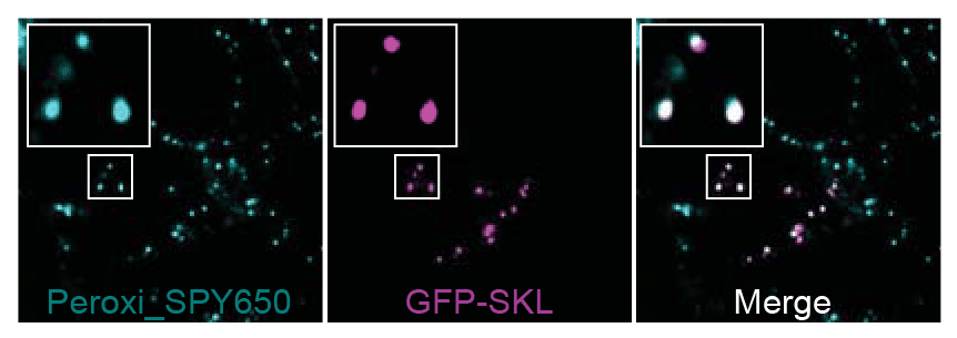
Figure 1: Peroxi_SPY650 GFP-SKL colocalisation.
The Power of Multicolour Imaging
Another exciting aspect of the Peroxi_SPY probes is the availability of two distinct fluorescence colours—Peroxi_SPY555 and Peroxi_SPY650—allowing researchers to use them in multicolour imaging experiments.
This feature is especially valuable when studying cellular processes involving multiple organelles or signalling pathways. With their high specificity and bright fluorescence, these probes provide clear, reproducible results without complex genetic modifications.
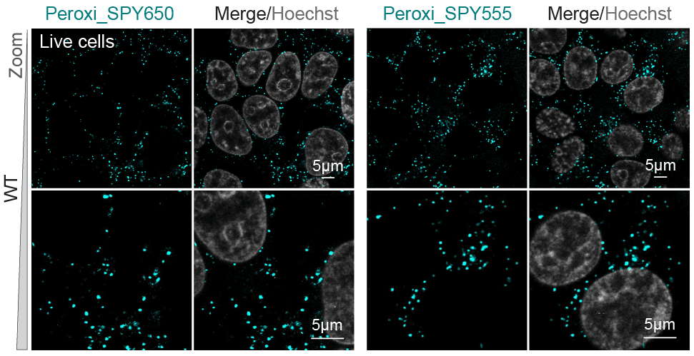
Figure 2: Peroxi _SPY555 and 650 HEK cells, hoechst.
Beyond Basic Research: Clinical and Diagnostic Applications
Peroxi_SPY probes are not just limited to basic research. They have the potential to significantly advance clinical diagnostics, particularly for diseases associated with peroxisomal dysfunction. Studies have demonstrated that these probes can effectively identify peroxisomal abnormalities in patient-derived cells and CRISPR/Cas9 models of disorders like Zellweger syndrome and Adrenoleukodystrophy.
This capability offers exciting possibilities for non-invasive diagnostic imaging, opening doors for more precise and efficient disease monitoring.
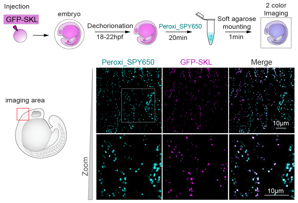
Figure 3: Peroxi_SPY650 Fish embryo.
The Bottom Line: Speed, Simplicity, and Reliability
If you're looking to improve your peroxisome imaging experiments, Peroxi_SPY probes represent a significant advancement. Their simple application, rapid staining protocol, and non-toxic nature make them ideal for a wide variety of live cell imaging needs. Whether for basic research or more specialised applications in clinical diagnostics, these probes provide an efficient, reliable solution that addresses many of the challenges researchers face in the lab today.
For researchers looking to push the boundaries of cellular imaging, Peroxi_SPY probes offer a powerful tool for clearer, faster, and more efficient peroxisome study.

Agnès Marcilly, MSc
Marketing Team at Tebubio
"With Peroxi_SPY probes, Spirochrome is simplifying peroxisome imaging, making it faster, more reliable, and accessible to researchers across diverse biological fields."
Name goes here
Position goes here
Description goes here
-
References
Figures 1, 2 and 3: Courtesy of Spirochrome AG, your trusted supplier for innovative imaging solutions.
Let’s Simplify Your Peroxisome Imaging
Looking for an easier, faster, and more reliable way to study peroxisomes? Contact our team to explore how Peroxi_SPY probes can streamline your research. Let’s discuss your needs and how we can support your imaging projects with a tailored solution.